Moms-to-be typically look forward to ultrasounds during pregnancy more than any other prenatal appointment. It’s easy to see why! It’s fun to get a sneak peek of your bundle of joy while your OBGYN looks for specific growth and development markers. Many women look forward to learning their baby’s sex as soon as possible and wait impatiently for their 20-week ultrasound. Others want to be surprised. Whether or not you intend to learn about your baby’s sex ASAP, it’s important to keep all your ultrasound appointments.
An ultrasound, also known as a sonogram, is typically performed by an ultrasound technician or sometimes by an OBGYN. It uses sound waves to create an image of the organs inside your body. (These sound waves are not harmful to you or your baby.) Ultrasounds during pregnancy help doctors diagnose many diseases and conditions, even those not related to pregnancy.
This article will cover why and how ultrasounds are used during your pregnancy, how many ultrasounds to expect while you’re pregnant, and what your OBGYN may be looking for at each ultrasound.
Most pregnant women typically only get two ultrasounds, one at the beginning of pregnancy and one about halfway through. Other women may have three or more ultrasounds done depending on a number of factors.
Your First Ultrasound
Your first ultrasound is called the “dating” or “viability” ultrasound. It’s typically done between 7 and 8 weeks to verify your due date, to look for a fetal heartbeat, and to measure the length of the baby from “crown to rump.” At this ultrasound, you’ll also learn whether you’re having one baby, pregnant with twins, or more! You may even get to see or hear your baby’s heartbeat during this appointment.
If you have irregular periods, or didn’t have a period after coming off birth control, this ultrasound will be especially helpful in determining a more accurate due date. Your due date is important because it helps your doctor know whether your baby’s development is on track each month.
We’ll perform this ultrasound at our Madison Women’s Health clinic.
What to Expect at Your First Ultrasound
When you’re just 7 or 8 weeks pregnant, your fetus is only about two centimeters long. In order to get a close enough view of your uterus and fetus, the dating ultrasound is done transvaginally. This means the ultrasound is done internally, literally “through the vagina.” A transvaginal ultrasound can be a little uncomfortable, but it is not painful. Most would say it feels less invasive than a gynecological exam that uses a speculum.
To perform this ultrasound, your OBGYN or ultrasound tech will gently insert a narrow ultrasound wand just inside your vagina. The transvaginal ultrasound wand is also called a transducer. It’s about three centimeters around, a little larger than a tampon. It will be covered by a condom and lubricant. The wand will not reach your cervix and is safe for your baby.
You may be asked to arrive at your first ultrasound with a full bladder. Having a fuller bladder helps to put your uterus in a better position for the ultrasound.
What Your Doctor is Looking for at Your First Ultrasound
- Viability of pregnancy
- Fetal heartbeat
- Fetal size
- Single or multiple pregnancy
Genetic Screening Ultrasound
If you choose to have prenatal genetic testing done, you’ll have your next ultrasound at 12 to 13 weeks gestation. This ultrasound is also called nuchal translucency screening. It’s offered to everyone and is covered by most insurance plans. This genetic screening ultrasound is optional.
During this ultrasound, your doctor will look for indicators of chromosomal disorders. Chromosomal disorders mean that the baby received an extra chromosome at conception and could have moderate to extreme physical or mental challenges. These disorders include:
- Trisomy 21, known as Down Syndrome
- Trisomy 13
- Trisomy 18
Read more about carrier screening and prenatal genetic testing. Also check out the article about at-home genetic testing kits.
What Your Doctor is Looking for at the Genetic Screening Ultrasound
This ultrasound will be an anatomical scan. Your doctor will look to see if all four limbs are present. They will also look for basic structures in the brain, the stomach, the bladder, the nasal bone, and last but not least, something called nuchal translucency. Nuchal translucency is a fluid sack at the back of the baby’s neck that is filled with lymphatic fluid. There are correlations between the size of that sack of fluid and the likelihood that the fetus could be affected by a major chromosomal disorder.
After the ultrasound has been performed, your OBGYN will interpret the results and share the information with you. You may also meet with a genetic counselor who could recommend having additional tests done to verify the ultrasound results.
Keep in mind that ultrasound screenings for other genetic disorders or anatomic abnormalities become more accurate further into the pregnancy.
We’ll perform this ultrasound at the Madison Women’s Health clinic.
Should you Get a Genetic Screening Ultrasound?
There’s no right or wrong answer to this question. Ultimately, the decision is up to you. Here are some good questions to ask yourself as you decide whether to have the genetic screening:
- Is there a family history of these genetic birth disorders?
- Would I terminate my pregnancy if there was a risk of Downs Syndrome, Trisomy 13, Trisomy 18, or other genetic disorder?
- Would knowing about my pregnancy’s risk of genetic defect make it easier to emotionally or physically prepare for a baby with a birth defect?
- Would it be easier for me to cope with and enjoy pregnancy if I focus on the more likely positive outcome rather than the chance of birth defects?
Whether you choose to have genetic screening done at this time is entirely your decision. Some women prefer to have as much information as possible as early as possible, while other women do not. If you’re still uncertain, you can discuss the pros and cons with your OBGYN.
Basic Anatomy Scan Ultrasound
This is the ultrasound that people look forward to the most! The full anatomy ultrasound is typically performed at about 20 weeks, or 5 months. As the name implies, this ultrasound will look at all the baby’s organ systems to make sure they’re present, are a normal size and shape, and are in the right location.
What to Expect at a Full Anatomy Scan Ultrasound
The full anatomy scan is a transabdominal ultrasound. It uses a transducer that looks a lot like a store checkout scanner. The ultrasound technician will put warm ultrasound gel on your stomach and then slide the transducer in the gel around your stomach. The gel helps the sound waves travel through your skin.
Tip: Come to your appointment with a relatively full bladder. This will make it easier for your ultrasound technician to get better images of your baby.
Because there are so many things to look for, this ultrasound will take at least 45 minutes—if your little one cooperates! If you’ve got an extra squirmy baby who is “camera shy,” it could take a few hours to get all the images that we need. Don’t worry, we have a lot of tricks to encourage your baby to change positions—everything from asking you to lay on one side and then the other, emptying your bladder or filling it, maybe even walking around. We’ll do whatever it takes to get the images we need to track your baby’s growth and development.
What Your OBGYN is Looking for at a Full Anatomy Scan
During the full anatomy, 20-week ultrasound, you can find out if your baby is male or female. If you want the sex to be a surprise, be sure to tell your technician know ahead of time so they don’t accidentally let it slip. When the scan is complete, Meriter will even send you a link to view some fun photos of your baby!
Your ultrasound technician will capture a large number images and measurements:
- limbs: arms, legs, feet, hands
- torso: chest, heart, kidneys, stomach, bladder, diaphragm, genitals
- head and face
- spine
- umbilical cord
- amount of amniotic fluid
- location, size, and shape of your placenta
- length of your cervix
After your ultrasound technician has captured all these images and measurements, your OBGYN will review the pictures and look for abnormalities such as congenital heart defects or cleft lip or palate. They’ll discuss their findings with you and help you understand what you’re looking at in the different images.
If everything looks normal and there are no other issues during your pregnancy, the next time you’ll see your baby is when he or she is in your arms! In the meantime, you can enjoy those 2D or 3D photos of your baby!
This ultrasound will be performed at UnityPoint Health – Meriter Hospital Center for Perinatal Care.
“Extra” Ultrasounds
Sometimes, women need additional ultrasounds during pregnancy. Your OBGYN may ask you to come in for additional ultrasounds to check your:
- Cervical length: if your cervix is shorter than expected, you may need to have your cervix checked regularly to be sure it stays closed so that you can maintain your pregnancy. If the cervix continues to shorten or thin, you may need a cerclage to help strengthen it until it’s time to deliver your baby. Cervical length ultrasounds occur at 16, 18, 20 and 22 weeks and are done transvaginally.
- Placental location and size: if your placenta is too small, if it is in an abnormal location or if it is an abnormal shape, then we will need to monitor it and the growth of your baby with regular ultrasounds. Your placenta is responsible for passing blood and nutrients to your baby, so it’s important thrat it is growing correctly.
You may need growth ultrasounds if you have:
- hypertension
- diabetes
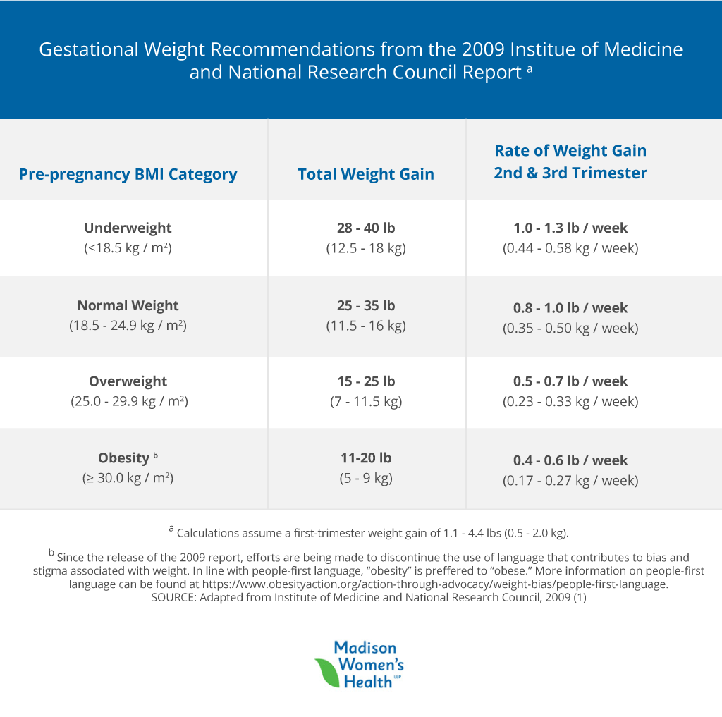
- high BMI (body mass index) going into pregnancy
- preeclampsia
- indicators that your placenta or uterus is not growing appropriately
Sometimes, growth ultrasounds are needed to check that your baby’s growth is continuing along the growth curve. They’re done at 28, 32, and 36 weeks. One way doctors estimate whether your baby is growing as expected is by measuring your fundal height. Fundal height is the number of centimeters from your pubic bone to the top of your uterus. This measurement typically increases about 1 cm each week. If your uterus has not grown appropriately in the last month, your OBGYN will surmise that your baby is also not growing and will want to perform monthly growth ultrasounds.
What to Expect at a Growth Ultrasound
These ultrasounds take less time than the full basic anatomy ultrasound because there are fewer measurements required. The ultrasound technician will measure the baby’s head circumference, bi-parietal diameter, abdominal circumference, and femur length.
What Your OBGYN is Looking for at Growth Ultrasounds
Your OBGYN is looking to see if your baby is staying on its growth curve. We will also use the measurements to estimate your baby’s weight. A large or extra large baby isn’t typically concerning. An extra-small baby or a baby who does not grow according to their growth curve could mean that the baby is not getting enough nourishment through the placenta and may need to be delivered early.
2D, 3D, and 4D Ultrasounds
2D ultrasounds are the black and white images that you’re probably used to seeing. To an untrained eye, they can look pretty fuzzy or obscure. However, they give the best definition of the structures of your growing peanut and are considered the “gold standard” of diagnostic imaging.
3D images are especially popular among parents-to-be who want to enjoy those cute baby pictures even before the baby is born! These pictures show facial features and look much more baby-like than the kind of obscure 2D images. 3D ultrasounds have usefulness beyond the cuteness factor, however! In the case of abnormalities of the spine or palate, 3D ultrasounds can help your OBGYN get a better idea of the severity.
4D images are like a 3D image, but show the baby moving around. They’re like getting to see a live action video of your little one. These are less commonly done because they don’t actually help with diagnoses. Depending on which perinatal center you go to, you might receive a link to view your ultrasound images or videos online.
While there are some stand-alone ultrasound centers offering to tell you your baby’s sex early on or to give you keepsake 3D or 4D images, these aren’t necessary and are rarely covered by insurance. You’ll find out everything you need to know during your appointments at the perinatal center — and those appointments will be covered by your insurance.
The best place to have an ultrasound performed is always at a clinic, where you will have access to a physician who has been trained in interpreting the images. At Madison Women’s Health, we’re happy to print off pictures for you to put in your baby’s scrapbook — or anywhere else you’d like to display those “coming soon” photos.
Final Thoughts
Ultrasounds during pregnancy are a fascinating way to get a glimpse of your developing baby. Don’t be afraid to ask your OBGYN for more details about genetic screening as you determine whether that is something you want done. And make sure you’ve set aside a good amount of time for your ultrasounds — especially the all-important full anatomy scan!
—
 Dr. Beth Wiedel has been providing healthcare to women in Madison since 2002 and is a founding partner of Madison Women’s Health. She shares the vision of all the partners of being a strong healthcare advocate for her patients, emphasizing compassion and communication throughout her practice.
 Dr. Ashley Durward has been providing healthcare to women in Madison since 2015 and joined Madison Women’s Health in 2019, specializing in high and low risk obstetrics, contraception and preconception counseling, management of abnormal uterine bleeding, pelvic floor disorders, and minimally invasive gynecologic surgery.


 Source:

